43 the human heart and its labels
How the Heart Works: Diagram, Anatomy, Blood Flow - MedicineNet The heart is located under the rib cage -- 2/3 of it is to the left of your breastbone (sternum) -- and between your lungs and above the diaphragm. The heart is about the size of a closed fist, weighs about 10.5 ounces, and is somewhat cone-shaped. It is covered by a sack termed the pericardium or pericardial sack. human system with label Human Male Skeleton - Stock Image - C024/9740 - Science Photo Library . Label The Human Stomach On A Diagram The Four Main Regions Of Stomach medicinebtg.com. stomach label anatomy diagram digestive human sphincter labeled regions system physiology identify curvatures its google tract body models types four
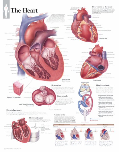
A Labeled Diagram of the Human Heart You Really Need to See The human heart, comprises four chambers: right atrium, left atrium, right ventricle and left ventricle. The two upper chambers are called the left and the right atria, and the two lower chambers are known as the left and the right ventricles. The two atria and ventricles are separated from each other by a muscle wall called 'septum'.

The human heart and its labels
File:Diagram of the human heart (cropped).svg - Wikipedia Added shadows. Left main pulmonary artery with its first division. 07:02, 2 June 2006: 650 × 650 (26 KB) Yaddah: Diagram of the human heart, created by Wapcaplet in Sodipodi. Cropped by ~~~ to remove white space (this cropping is not the same as Wapcaplet's original crop). == See also == * Image:Diagram of the human heart.svg - original How to Draw a Human Heart: 11 Steps (with Pictures) - wikiHow Label the parts of the heart if you'd to reference it for anatomy. If you're trying to identify parts of the heart for a class you're taking, it's good practice to draw the heart yourself and label each segment. You can refer to your textbook in order to label the: [9] Aorta Superior vena cava Inferior vena cava Right and left atria The Anatomy of the Heart, Its Structures, and Functions - ThoughtCo The heart is the organ that helps supply blood and oxygen to all parts of the body. It is divided by a partition (or septum) into two halves. The halves are, in turn, divided into four chambers. The heart is situated within the chest cavity and surrounded by a fluid-filled sac called the pericardium. This amazing muscle produces electrical ...
The human heart and its labels. 147 Heart Anatomy With Labels Premium High Res Photos - Getty Images Browse 147 heart anatomy with labels stock photos and images available, or start a new search to explore more stock photos and images. of 3. NEXT. parts of the heart label Label The Heart Diagram - Human Anatomy tartrerepub.blogspot.com. heart diagram worksheet blank label worksheets sparklebox human anatomy related. Lab 2 Pig Heart-Labeled | Cardiovascular, Estudos . heart anatomy lab pig physiology human labeled dissection sheep posterior pigs body salvo escolha pasta kctcs bluegrass district edu. Heart: Anatomy and Function - Cleveland Clinic Your heart is the primary organ of your circulatory system. It pumps blood throughout your body, controls your heart rate and maintains blood pressure. Your heart is a bit like a house. It has walls, rooms, doors, plumbing and an electrical system. All the parts of your heart work together to keep blood flowing and send nutrients to your other ... A Diagram of the Heart and Its Functioning Explained in Detail The heart blood flow diagram (flowchart) given below will help you to understand the pathway of blood through the heart.Initial five points denotes impure or deoxygenated blood and the last five points denotes pure or oxygenated blood. 1.Different Parts of the Body ↓ 2.Major Veins ↓ 3.Right Atrium ↓ 4.Right Ventricle ↓ 5.Pulmonary Artery ↓ 6.Lungs
Human Heart Diagram - Human Body Pictures - Science for Kids Find free pictures, photos, diagrams, images and information related to the human body right here at Science Kids. Photo name: Human Heart Diagram Picture category: Human Body Image size: 70 KB Dimensions: 600 x 600 Photo description: This is an excellent human heart diagram which uses different colors to show different parts and also labels a number of important heart component such as the ... Heart - Wikipedia The human heart is situated in the mediastinum, at the level of thoracic vertebrae T5 - T8. A double-membraned sac called the pericardium surrounds the heart and attaches to the mediastinum. [15] The back surface of the heart lies near the vertebral column, and the front surface sits behind the sternum and rib cartilages. [7] Heart Labeling Quiz: How Much You Know About Heart Labeling? Here is a Heart labeling quiz for you. The human heart is a vital organ for every human. The more healthy your heart is, the longer the chances you have of surviving, so you better take care of it. Take the following quiz to know how much you know about your heart. Questions and Answers. 1. 13 parts of the human heart (and its functions) - LORECENTRAL Its opening (generated by the systole of the atrium) causes blood to travel between both regions. 3. Left Ventricle. Another major part of the heart. The left ventricle receives oxygen-rich blood from the left atrium and sends it to the rest of the body through the aortic artery. 4. Aortic sigmoid valve.
PDF Analyzing the Human Heart - Beyond the Classroom working. Its job is to pump blood to the lungs and to all of the body tissues. In this activity you will use a diagram of the heart to analyze the way in which the heart works. l. Using the following word list, label the various parts of the heart on the diagram. Right ventricle Left venfficle Upper vena cava Lower vena cava Aorta Heart: illustrated anatomy - e-Anatomy - IMAIOS This interactive atlas of human heart anatomy is based on medical illustrations and cadaver photography. The user can show or hide the anatomical labels which provide a useful tool to create illustrations perfectly adapted for teaching. Anatomy of the heart: anatomical illustrations and structures, 3D model and photographs of dissection. Heart Diagram - 15+ Free Printable Word, Excel, EPS, PSD Template ... Teachers and students use the heart diagram, in biological science, to study the structure and functions of a human being's heart. ... Label The Parts Of The Heart. depts.washington.edu | Having the heart diagram for studies or for scientific purpose has been made easy through this template. It shows a heart picture with all its parts labeled ... File:Diagram of the human heart (cropped).svg - Wikimedia Aug 08, 2022 · Add Inferior vena cava and pericardium labels: 18:08, 14 August 2018: 656 × 631 (209 KB) ... Diagram of the human heart, created by Wapcaplet in Sodipodi. Cropped by ...

Congestive Heart Failure: The Essence of Heart Failure Course | CEUfast Nursing Continuing Education
How the Heart Works - The Heart | NHLBI, NIH - National Institutes of ... The Heart. The heart is an organ about the size of your fist that pumps blood through your body. It is made up of multiple layers of tissue. Your heart is at the center of your circulatory system. This system is a network of blood vessels, such as arteries, veins, and capillaries, that carries blood to and from all areas of your body.

Simplified Heart Labeled Decal | Shop Fathead Anatomical Images Graphics
Labelling the heart — Science Learning Hub Labelling the heart — Science Learning Hub Labelling the heart Add to collection The heart is a muscular organ that pumps blood through the blood vessels of the circulatory system. Blood transports oxygen and nutrients to the body. It is also involved in the removal of metabolic wastes. Topics Concepts Citizen science Teacher PLD Glossary Sign in

Human Nervous System Structure and Functions Explained With Diagrams - Bodytomy
File:Diagram of the human heart (no labels).svg - Wikimedia File:Diagram of the human heart (no labels).svg. From Wikimedia Commons, the free media repository. File. File history. File usage on Commons. Metadata. Size of this PNG preview of this SVG file: 498 × 599 pixels. Other resolutions: 199 × 240 pixels | 399 × 480 pixels | 639 × 768 pixels | 851 × 1,024 pixels | 1,703 × 2,048 pixels | 533 × ...

Human Heart - Diagram and Anatomy of the Heart - Innerbody The heart contains 4 chambers: the right atrium, left atrium, right ventricle, and left ventricle. The atria are smaller than the ventricles and have thinner, less muscular walls than the ventricles. The atria act as receiving chambers for blood, so they are connected to the veins that carry blood to the heart.

New Photos in Anatomy of human body organs
Heart Diagram with Labels and Detailed Explanation - BYJUS Diagram of Heart. The human heart is the most crucial organ of the human body. It pumps blood from the heart to different parts of the body and back to the heart. The most common heart attack symptoms or warning signs are chest pain, breathlessness, nausea, sweating etc. The diagram of heart is beneficial for Class 10 and 12 and is frequently ...

Your New Puppy Author Cindy Moore, cindy@k9web.com Copyright 1992-99. Table of Contents ...
Human Heart (Anatomy): Diagram, Function, Chambers, Location ... Heart Treatments. Exercise: Regular exercise is important for heart health and most heart conditions.Talk to your doctor before starting an exercise program if you have heart problems. Angioplasty ...

Business Diary: October 2011
Label the heart — Science Learning Hub Label the heart Interactive Add to collection In this interactive, you can label parts of the human heart. Drag and drop the text labels onto the boxes next to the diagram. Selecting or hovering over a box will highlight each area in the diagram. Pulmonary artery Left ventricle Right ventricle Left atrium Right atrium Pulmonary vein Semilunar valve
The Heart | Scientific Publishing
Dare (album) - Wikipedia Dare earned considerable income for record labels Virgin and A&M; in Virgin's case, it gave the label the first chart-topping album since Mike Oldfield's Tubular Bells in 1973. "Don't You Want Me" was the label's first ever chart-topping single. The success of Dare was responsible for saving the label from impending bankruptcy.
Sampoerna Wallpaper: The Heart Diagram Labeled
Parts Of The Human Heart | Science Trends The parts of the human heart can be broken down into four chambers, muscular walls, vessels, and a conductive system. The two upper chambers are called the atria, with lower parts called ventricles. These all work together to make up the vital function of your heart. Everybody knows that the human heart is the essential organ in our bodies.
Post a Comment for "43 the human heart and its labels"