39 external structure of the heart with labels
Human Heart (Anatomy): Diagram, Function, Chambers ... The heart is a muscular organ about the size of a fist, located just behind and slightly left of the breastbone. The heart pumps blood through the network of arteries and veins called the ... The Tenors - Wikipedia The Tenors (formerly known as The Canadian Tenors) are a vocal group consisting of Victor Micallef, Fraser Walters, and Clifton Murray.They perform operatic pop music that is a mixture of classical and pop, featuring songs such as "The Prayer", Panis angelicus, and Leonard Cohen's Hallelujah.
40.3A: Structures of the Heart - Biology LibreTexts Structure of the Heart. The heart is a complex muscle that pumps blood through the three divisions of the circulatory system: the coronary (vessels that serve the heart), pulmonary (heart and lungs), and systemic (systems of the body). Coronary circulation intrinsic to the heart takes blood directly from the main artery (aorta) coming from the ...
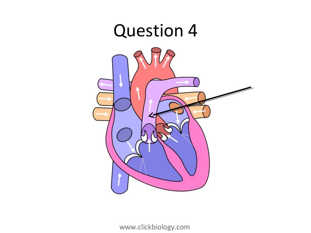
External structure of the heart with labels
Lesson | The Heart - External Structure | Encounter Edu In this lesson students begin their exploration of the circulatory system, labelling a diagram of the external structures and identifying arteries and veins. They will go on to explain where blood enters and leaves the heart. Learning outcomes Human Heart Diagram Labeled - Science Trends The endocardium is the inner portion of the outer wall, and the endocardium is what contacts the blood in the heart. The heart's atrioventricular valves are structures that join the atria and ventricles of the heart together. This group of valves is comprised of the tricuspid valve and the mitral valve. Chapter 19: The Heart Flashcards | Quizlet •Allows heart to beat without friction, gives it room to expand and resists excessive expansion •Parietal pericardium-tough outer, fibrous layer of connective tissue-inner serous layer •Visceral pericardium (a.k.a. epicardium of heart wall)-serous lining of sac turns inward at base of heart to cover the heart surface
External structure of the heart with labels. Heart - External Features - Anatomy QA Apex beat. Is the lowermost and outermost thrust of the heart, felt on the front of the chest. In adults it is felt in the left 5 th intercostal space 9cm. from the median plane (just medial to the midclavicular line). In infants it is felt in the 3 rd intercostal space just lateral to the midclavicular line.. Dextrocardia. It is a congenital anomaly in which the heart lies on the right side ... Layers of the heart: Epicardium, myocardium, endocardium ... The heart is a muscular organ found in the middle mediastinum that pumps blood throughout the body. It is housed in the pericardial sac, which protects it and assists with its mechanics. Recalling from the heart anatomy, it has two atria and two ventricles that make up elements and important steps for the heart cycle. External anterior heart labeling Quiz - PurposeGames.com About this Quiz. This is an online quiz called External anterior heart labeling. There is a printable worksheet available for download here so you can take the quiz with pen and paper. Heart Anatomy: Heart Dissection The letters indicated in the text refer to the labels on the picture. The anterior surface of the heart is characterized by the presence of the large arteries leaving the base of the heart, the pulmonary trunk (H) and the aorta (G). The pulmonary trunk is the vessel that divides to give rise to the two pulmonary arteries going to each lung.
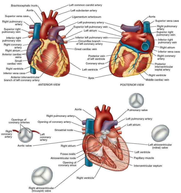
Solved Art-Labeling Activity: Overview of the external ... art-labeling activity: overview of the external anatomy of the heart anterior view res great cardiac vein aortic arch right coronary artery left coronary artery left pulmonary veins ascending aorta left pulmonary artery anterior interventricular artery superior vena cava pulmonary trunk auricle of left atrium circumflex artery auricle of right … A Labeled Diagram of the Human Heart You Really Need to ... The human heart, comprises four chambers: right atrium, left atrium, right ventricle and left ventricle. The two upper chambers are called the left and the right atria, and the two lower chambers are known as the left and the right ventricles. The two atria and ventricles are separated from each other by a muscle wall called 'septum'. Chapter 22 Heart Flashcards | Quizlet Label the coronary arteries in an anterior view of the heart. Label the order that blood flows through in the heart, using the arrows as guides. Label the components of the heart wall. Label the components of the heart as seen from a posterior view. Label the major coronary veins. Label the components of the conduction system. Heart Anatomy: size, location, coverings and layers ... The heart wall is composed of three layers: the epicardium, myocardium, and endocardium. Location of the heart in the mediastinum. The superficial epicardium is the visceral layer of the serous pericardium. The middle layer is the myocardium and is composed mainly of cardiac muscle and forms the bulk of the heart.
Heart Anatomy: Labeled Diagram, Structures, Blood Flow ... Let's begin with the chambers of the heart. There are 4 chambers, labeled 1-4 on the diagram below. To help simplify things, we can convert the heart into a square. We will then divide that square into 4 different boxes which will represent the 4 chambers of the heart. Label the heart - Science Learning Hub In this interactive, you can label parts of the human heart. Drag and drop the text labels onto the boxes next to the diagram. Selecting or hovering over a box will highlight each area in the diagram. Pulmonary vein Right atrium Semilunar valve Left ventricle Vena cava Right ventricle Pulmonary artery Aorta Left atrium Download Exercise Tweet Heart Diagram with Labels and Detailed Explanation - BYJUS Diagram of Heart. The human heart is the most crucial organ of the human body. It pumps blood from the heart to different parts of the body and back to the heart. The most common heart attack symptoms or warning signs are chest pain, breathlessness, nausea, sweating etc. The diagram of heart is beneficial for Class 10 and 12 and is frequently ... Ch. 19 Circulatory System- heart Flashcards | Quizlet Correctly label the external anatomy of the anterior heart. Place the labels in order denoting the flow of blood through the pulmonary circuit beginning with the right atrium and ending in the left atrioventricular valve. The first and last structures are given. Right atrium 1. tricuspid valve 2. right ventricle 3. pulmonary valve
Heart Structure and Function
The Anatomy of the Heart, Its Structures, and Functions The heart is the organ that helps supply blood and oxygen to all parts of the body. It is divided by a partition (or septum) into two halves. The halves are, in turn, divided into four chambers. The heart is situated within the chest cavity and surrounded by a fluid-filled sac called the pericardium. This amazing muscle produces electrical ...

Medical Tourism News and Views: How does the heart work?
Heart Anatomy Labeling Game This is an online quiz called Heart Anatomy Labeling Game. There is a printable worksheet available for download here so you can take the quiz with pen and paper. Your Skills & Rank. Total Points. 0. Get started! Today's Rank--0. Today 's Points. One of us! Game Points. 19. You need to get 100% to score the 19 points available.
heart
Dapagliflozin - Wikipedia Dapagliflozin, sold under the brand names Farxiga (US) and Forxiga (EU) among others, is a medication used to treat type 2 diabetes. It is also used to treat adults with certain kinds of heart failure and chronic kidney disease.
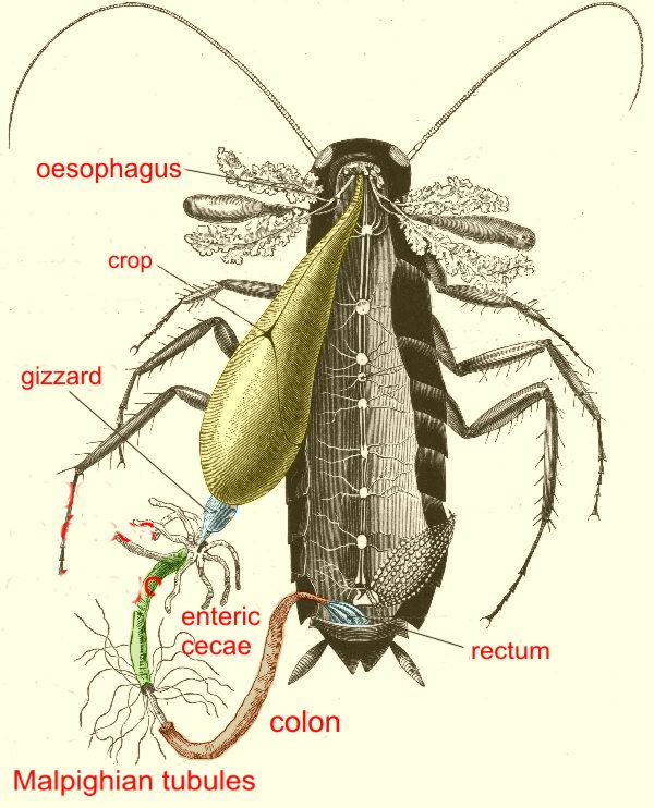
cockroaches parasites 3
The 3 Layers of the Heart Wall - ThoughtCo The heart is an extraordinary organ. It is about the size of a clenched fist, weighs about 10.5 ounces and is shaped like a cone. Along with the circulatory system, the heart works to supply blood and oxygen to all parts of the body. The heart is located in the chest cavity just posterior to the breastbone, between the lungs, and superior to the diaphragm.

Ongzi's Lifelong Learning: Typhoid Fever
Heart Anatomy | Anatomy and Physiology - Lumen Learning The wall of the heart is composed of three layers of unequal thickness. From superficial to deep, these are the epicardium, the myocardium, and the endocardium. The outermost layer of the wall of the heart is also the innermost layer of the pericardium, the epicardium, or the visceral pericardium discussed earlier. Figure 6.
Congestive Heart Failure: The Essence of Heart Failure Course | CEUfast Nursing Continuing Education
The structure of the heart - Structure and function of the ... Each side of the heart consists of an atrium and a ventricle which are two connected chambers. The atria (plural of atrium) are where the blood collects when it enters the heart. The ventricles...

Anatomy Heart Quizlet
PDF Anatomy of Heart Labeled and Unlabeled Images (a) Anterior view of the external heart C' 2019 Pearson Education. Aort'c arch Ligamentum arteriosum Left pulmonary artery Left pulmonary ve ns Auricle of left atrium Circumflex artery Left coronary artery (in atrioventricular sulcus) Great cardiac vein Left ventricle Anterior interventricular artery (in anterior interventricular sulcus) Apex

The Heart - Biology Student
Structure of the Heart | SEER Training The human heart is a four-chambered muscular organ, shaped and sized roughly like a man's closed fist with two-thirds of the mass to the left of midline. The heart is enclosed in a pericardial sac that is lined with the parietal layers of a serous membrane. The visceral layer of the serous membrane forms the epicardium. Layers of the Heart Wall

DNA Replication - Structure - Stages of Replication - TeachMePhyiology
Diagram of the human heart Images, Stock ... - Shutterstock Diagram of the human heart royalty-free images. 14,495 diagram of the human heart stock photos, vectors, and illustrations are available royalty-free. See diagram of the human heart stock video clips. Image type.
HeartComplete
Label the Heart - The Biology Corner Shows a picture of a heart with letters and blanks for practice with labeling the parts of the heart and tracing the flow of blood within the heart.

Pick Correct Options For Human Heart Quiz - ProProfs Quiz
Solved -labeling Activity: External Anatomy of the Sheep ... Anatomy and Physiology. Anatomy and Physiology questions and answers. -labeling Activity: External Anatomy of the Sheep Heart Part A Drag the labels to the appropriate location in the figure. Reset Help Lolt ventric Pulmonary trunk Lolt atrium Lohtaude Right trum Posterior Interventricular sules = Pulmonary veins Art Right vorticle Anterior ...

The Heart | S-cool, the revision website
Flat vs. Deep Website Hierarchies - Nielsen Norman Group Nov 10, 2013 · Left: a flat site hierarchy, with few vertical levels. Right: a deep site hierarchy has the same information organized into more sublevels. Both of these site hierarchies start at the top with a single homepage, but the information below that page is organized quite differently: the website on the left has 8 major categories, but the site on the right has only 4.

Pin on Anatomy Human Other Bambie Reed
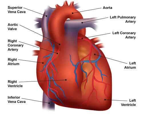
Human Heart - Anatomy, Functions and Facts about Heart The external structure of the heart has many blood vessels that form a network, with other major vessels emerging from within the structure. The blood vessels typically comprise the following: Veins supply deoxygenated blood to the heart via inferior and superior vena cava, and it eventually drains into the right atrium.
Heart - Labelled diagram
Structure Of The Heart | A-Level Biology Revision Notes The heart is a hollow muscular organ that lies in the middle of the chest cavity. It is enclosed in the pericardium, which protects the heart and facilitates its pumping action. The heart is divided into four chambers: The two atria (auricles): these are the upper two chambers. They have thin walls which receive blood from veins.

Labeling the Heart
Internal Structure of the Heart | Contemporary Health Issues It is marked by the presence of four openings that allow blood to move from the atria into the ventricles and from the ventricles into the pulmonary trunk and aorta. Located in each of these openings between the atria and ventricles is a valve, a specialized structure that ensures one-way flow of blood.

Heart anatomy vector illustration - VectorMine
Gadgets - TechCrunch Cultivated meat, grown in a bioreactor rather than out on the range, might be one of the big food trends of the decade. But it’s relying on tech built around multiplying yeast and bacteria cells
.jpg)
Heart Model & Sheep Heart Practice Quiz - ProProfs Quiz
Chapter 19: The Heart Flashcards | Quizlet •Allows heart to beat without friction, gives it room to expand and resists excessive expansion •Parietal pericardium-tough outer, fibrous layer of connective tissue-inner serous layer •Visceral pericardium (a.k.a. epicardium of heart wall)-serous lining of sac turns inward at base of heart to cover the heart surface
Post a Comment for "39 external structure of the heart with labels"